
Dr. Hai Song’s group collaborated with Dr. Kingston Mak’s group and published a research article on Nature communications entitled with “Reciprocal inhibition of YAP/TAZ and NF-κB regulates osteoarthritic cartilage degradation” on Nov. 1st 2018.
Osteoarthritis (OA) is one of the most common degenerative diseases and the incidence increases significantly with age. The disease is characterized by progressive degradation of articular cartilage, subchondral bone thickening and osteophyte formation, which ultimately leads to loss of joint mobility and joint functions. However, the molecular mechanisms regulating these processes in articular chondrocytes remain unclear. These regulatory processes are highly relevant to the onsets, pathogenesis and progression of OA.
Hippo signaling is identified to control organ size and tissue regeneration in many organs. However, whether the Hippo-YAP/TAZ pathway plays a role in regulating inflammatory response during OA pathogenesis remains elusive. The authors found that articular cartilage with OA showed significant reduction of YAP expression in mouse OA model. Furthermore, YAP expression levels were concomitantly reduced according to the severity and OARSI grade of OA in human patient samples
To elucidate the functional role of YAP in OA pathogenesis in vivo, the authors first generated Col2a1-Yap1tg/+ transgenic mice. Under osteoarthritic condition, Col2a1-Yap1tg/+ transgenic mice displayed remarkably better cartilage integrity than that of the wild-type mice. In addition,TNFα or IL1β treatment in transgenic chondrocytes led to lower induction of matrix degrading enzymes, but higher expression of cartilage ECM and YAP target genes than those of the wild-type chondrocytes. However under OA condition, cartilage degradation was significantly more severe in Yap1f/f;Col2a1-Cre mutant mice than that of the control mice. Mechanistically, inflammatory cytokines, such as TNFα or IL-1β inhibits the expression of Yap1/Taz and their target genes Ctgf and Cyr61. Besides, Gal4/TEAD4-luciferase reporter activity was inhibited in response to TNFα treatment in a dose dependent manner in primary chondrocytes. Furthermore, the inflammatory cytokines triggered the Hippo signaling activation and YAP degradation. The authors found that the protein kinase TAK1 was involved in the regulation of YAP phosphorylation and proteasome-medicated degradation. On the other hand, the authors found that YAP inhibits TAK1 substrate accessibility, abrogates IKKα/β activation and suppresses IκBα degradation, and p65 activation and nuclear translocation, which results in attenuation of NF-κB signaling activity and osteoarthritic cartilage degradation. These findings suggest that targeting YAP is a viable strategy for treating OA.
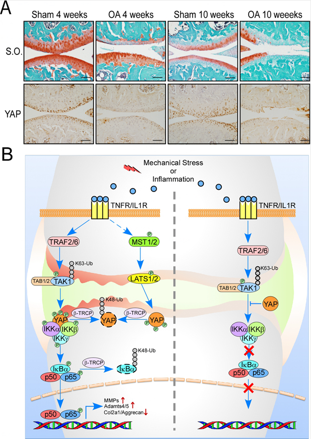
Figure A. YAP expression level is greatly downregulated in articular cartilage in OA mice. Figure B. A model of reciprocal antagonism between Hippo-YAP/TAZ and NF-κB pathway.
The project was completed by Hai Song’s Lab at Zhejiang University and Kingston Mak’s lab at the Chinese University of Hong Kong. The postdoc fellow Yujie Deng in Hai Song’s group is the first author. Prof. Hai Song and Dr. Kingston Mak are the corresponding authors. This work was supported by the National Natural Science Foundation of China (grant number 31471368) and the Zhejiang Provincial Natural Science Foundation of China (grant number LR16C120001) to H.S., and the National Natural Science Foundation of China (grant number 81700002) and the International Postdoctoral Research Fellowship Program to Y.D., and the Seed Fund of the School of Biomedical Sciences, The Chinese University of Hong Kong (4620504) to K.M. H.S. is a scholar in the National 1000 Young Talents Program.
Links: https://www.nature.com/articles/s41467-018-07022-2

